

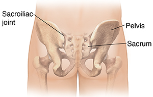
The Sacroiliac (SI) joint is a synovial (diarthrodial) joint that connects bilaterally ilium to central sacrum forming left & right SI joint in the tailbone. The joint is supported by multiple ligaments & muscles which contribute to pelvic stability & force transfer. It serves as shock absorbers for the pelvis and low back. The SI joints move constantly when the body is in motion, provide stability and structural support to the lower part of the body.
Painful SI conditions can be traumatic, infectious, metabolic, inflammatory, neoplastic, degenerative. Nonbacterial, acute sacroiliitis can be associated with ankylosing spondylitis, gout, RA, psoriasis trauma. The most common painful SIJ presentation seen in a clinic is a mechanical lesion associated trauma, as well as adjacent lumbar arthropathy and/or fusion.
The other most common cause is degenerative arthritis of the joint. Stretching, straining, and tearing of the primary SI joint ligaments then cause weakening and abnormal motion of the joints, resulting in painful ligaments and joints as well as spasm.
Symptoms
Patients complain of pain catching and increased pain with lower extremity loading, such as unimodal standing or landing and propulsion with gait.
Parasthesias, numbness or weakness is not commonly associated with painful mechanical SIJ states. Most patients experience low back pain that is worsened by sitting, standing, and bending at the waist. Frequent changes in posture are needed. Muscle insertions near the area, such as gluteus maximus & hamstrings refer pain to the hip & ischial area, respectively, when stressed
Diagnosis
Identifying SI joint dysfunction requires obtaining a detailed medical history, analyzing the mechanism of injury, and conducting a comprehensive examination of the spine and pelvis. SIJ evaluation also includes manual provocation tests & diagnostic injections.
Not all patients show obvious signs of SI joint dysfunction, and X-rays, MRIs, CT scans, and bone scans of the pelvis will generally be normal.
Management
- Education: proper ergonomic training for bending, lifting, and stretching. Rehabilitation techniques (flexibility, core strengthening/stabilization, and joint manipulation)
- Physical modalities like deep heat, TENS, mobilization
- Exercises for strengthening & posture enhancement.
- Injection of Steroid and analgesic provide best option for quickly reducing inflammation along the joint line.
- Prolotherapy the strength of the SI joint ligaments and helps restore normal motion. It is very good for resistant or chronically recurring cases.
- Surgical fusion of the SI joint (a last resort, performed only in cases of severe debilitating instability)
Other diseases which can be treated at our pain clinic-
1. Other joint pain:
- Frozen shoulder
- Tennis elbow
- Golfers elbow
- Osteoarthritis
- Sprain in joints
- Bursitis
- Tendinopathies
2. Sports injury
3. Rheumatoid arthritis
4. Gout
5. Atypical face pain
6. Pain in Middle back (Thoracic region)
7. Myofacial / Musculoskeletal Pain Syndrome
8. Any persistent pain at 6 weeks of Medical and/Or Surgical treatment.